
Description: Explore how silicon Micro-OLEDs, 3D-LUT calibration, and DICOM Part 14 GSDF compliance enable medical endoscopic displays to achieve ultra-high fidelity and sub-mm surgical precision.
Traditional open surgical procedures rely heavily on the unaided visual judgement of highly experienced surgeons. Today, advanced technologies are transforming surgical practice across the medical industry.
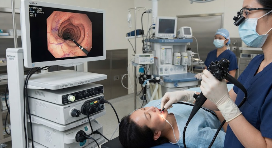
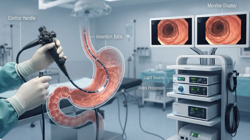
Medical endoscopic micro-display modules are widely deployed in minimally invasive surgical head-mounted displays, 3D electronic endoscope eyepieces, and fluorescence-guided surgical terminals. For instance, minimally invasive approaches are routinely used for sub-1 cm thyroidectomies to remove thyroid cancer via trans-axillary access.
Another representative procedure is laparoscopic liver resection with ICG fluorescence guidance. The liver hosts an extremely dense, complex vascular network; conventional open liver resection entails substantial tissue trauma and significant blood loss. With minimally invasive laparoscopic techniques, surgeons can precisely demarcate tumour margins, deep microvessels and bile ducts within confined operative fields. Following intravenous injection of indocyanine-green (ICG) fluorescent dye, tumour lesions emit fluorescence under near-infrared illumination. Leveraging high dynamic contrast and an ultra-wide colour gamut (>98% DCI-P3), micro-display modules render the saturated fluorescent-green tumour boundary accurately overlaid against the haemoglobin-rich red tones of healthy liver parenchyma. Surgeons can identify micro-metastatic foci as small as 0.5 mm and avoid inadvertent injury to major blood vessels.
When wearing head-mounted hardware, operating surgeons observe ultra-sharp imagery with clinically validated colour fidelity directly from the endoscopic feed, substantially reducing physical and cognitive workload. Many industry practitioners wonder how medical endoscopic micro-display modules achieve such high-resolution performance and meet strict clinical colour-rendering requirements. This article provides a detailed technical breakdown.
1. Ultra-Dense Silicon-Based CMOS Backplanes with Pixel-Level Isolation
Medical-grade endoscopic micro-display modules adopt 0.39-inch to 0.7-inch silicon-based Micro-OLED panels to deliver ultra-high pixel density, eliminating visible screen-door effects and supporting precise surgical navigation. Deep-trench isolation (DTI) structures are implemented within the CMOS pixel array to realise exceptional contrast ratios. Sharp edge definition across light-to-dark transitions enables clinicians to distinguish tissue boundaries reliably.
- Benchmark Case: Olympus VISERA ELITE III 3D Module (Olympus 3D Surgical Electronic Endoscopy System)
- Application: Gastrointestinal, respiratory and general minimally invasive endoscopic visualisation.
- Implementation: Its 3D-assisted viewing and eyepiece assembly integrates silicon-based display units exceeding 3000 PPI. Combined with DTI isolation technology, the system reveals tiny early-malignant mucosal elevations while visualising intricate microvascular networks.
2. Clinical Colour-Gamut Calibration for Vascular and Soft-Tissue Visualisation (R9 Index & 3D-LUT)
Minimally invasive surgery demands reliable differentiation of microvessels, adipose tissue, muscle and mucosal membranes. Medical endoscopic micro-display modules must cover most of the DCI-P3 colour gamut with special tuning for saturated red reproduction (R9 index) to minimise overall colour error. 3D colour-lookup-tables (3D-LUTs) are embedded within driver-IC silicon to faithfully reproduce in-vivo tissue appearance under cold-light illumination (LED / xenon sources) or ICG fluorescence excitation.
- Benchmark Case: Karl Storz IMAGE1 S™ Rubina / 3D (Karl Storz 3D Fluorescence Endoscopy System)
- Application: ICG near-infrared fluorescence tumour-margin identification and 3D laparoscopic surgery.
- Implementation: The 3D display and eyepiece receiver module delivers >98% DCI-P3 coverage alongside high dynamic contrast. During tumour-resection procedures, outstanding colour accuracy clearly separates saturated fluorescent-green lesions from native red-toned healthy tissue, supporting confident intra-operative decision-making.
3. Low-Light Detail Rendering and DICOM Part 14 Greyscale Compliance
Endoscopic fields-of-view frequently include deep shadowed cavities, imposing stringent low-light performance requirements. Module driver ICs implement 10-bit / 12-bit PWM dimming alongside reconstructed low-greyscale gamma curves to suppress banding and noise artefacts in dark regions. Firmware implements the DICOM Part 14 (GSDF) medical-imaging standard greyscale response function, ensuring consistent lesion-contrast representation regardless of the viewing hardware.
- Benchmark Case: Intuitive Surgical da Vinci Xi / SP Surgeon-Console System
- Application: Complex minimally invasive general, urological and gynaecological robotic surgery.
- Implementation: The console’s stereo viewer integrates dual high-PPI silicon-based micro-display modules. Hardware implements DICOM Part 14-aligned greyscale response together with factory-calibrated binocular gamma matching. Even under dim operative lighting, fine anatomical features such as microsurgical sutures and tiny microvessels are reproduced without distortion to enhance procedural safety.
4. Microsecond-Scale Photometric Response and Ultra-Low-Latency Refresh Rates
Capitalising on silicon micro-displays’ ultrafast electro-optical response paired with 90 Hz–120 Hz refresh rates, motion smear is effectively eliminated. During rapid probe manipulation and other high-frequency surgical manoeuvres, imagery updates without perceptible lag or ghosting. This mitigates operator-induced vestibular discomfort and reduces cumulative fatigue.
- Benchmark Case: Sony HMS-3000MT (Sony 3D Medical Head-Mounted Display System)
- Application: Laparoscopic and thoracoscopic minimally invasive surgery.
- Implementation: Independent high-resolution OLED micro-display panels serve each eye. Microsecond-scale luminescence response eliminates smear during rapid endoscope movements. Spectral optimisation targeting the red spectral band (R9 index) assists surgeons in discriminating blood vessels, fat and mucosal tissue.
5. Chip-Level Binocular Calibration and Hardware-Accelerated Distortion Compensation
Prior to factory release, luminance, colour temperature (e.g. 6500 K / 9300 K) and gamma parameters for left- and right-channel displays are written to on-board EEPROM memory, eliminating binocular visual mismatch that can trigger operator dizziness. Distortion and chromatic-aberration compensation lookup tables (LUTs) are implemented directly within the micro-display driver ASIC.
- Benchmark Case: Augmedics Xvision Spine System (FDA-Cleared AR-Guided Spinal Endoscopic Navigation Headset)
- Application: Minimally invasive spinal endoscopy and orthopaedic anatomical navigation.
- Implementation: Custom high-brightness, low-latency silicon-based Micro-OLED micro-display modules are deployed. Distortion and chromatic-aberration compensation LUTs reside within the micro-display driver ASIC hardware layer. CT scans, endoscopic video feeds and real-patient spinal anatomy are overlaid with minimal latency and precise spatial registration, reducing surgeon visual fatigue.
Authoritative References
[1] Society of American Gastrointestinal and Endoscopic Surgeons (SAGES). Guidelines and Technology Reviews for Minimally Invasive and Laparoscopic Surgery.
[2] IEEE Transactions on Medical Imaging. Fluorescence-Guided Surgery and High-Gamut DCI-P3 Color Mapping in Near-Infrared Endoscopy.
[3] International Commission on Illumination (CIE). CIE 013.3-1995: Method of Measuring and Specifying Colour Rendering Properties of Light Sources (R9 Index Evaluation).
[4] SPIE Digital Library. Optical System Design and Human Ergonomics for Surgical Head-Mounted Near-Eye Displays.
[5] Olympus Corporation. VISERA ELITE III 3D Surgical Electronic Endoscopy System Technical Architecture Specification.
[6] Karl Storz SE & Co. KG. IMAGE1 S™ Rubina™ Near-Infrared Fluorescence Imaging System Technical Whitepaper.
[7] Intuitive Surgical, Inc. da Vinci Xi/SP Surgical System Endoscopic Vision Subsystem Technical Overview.
[8] National Electrical Manufacturers Association (NEMA) / DICOM Standards Committee. DICOM PS3.14: Digital Imaging and Communications in Medicine – Part 14: Grayscale Standard Display Function (GSDF).
[9] Sony Corporation. Sony HMS-3000MT 3D Medical Head-Mounted Display Technical Specification Manual.
[10] U.S. Food and Drug Administration (FDA). Augmedics xvision-Spine System (XVS) 510(k) Regulatory Clearance Documentation (K192025).
Legal Disclaimer
This white-paper is intended solely for technical reference and does not constitute clinical guidance, engineering consultancy, procurement advice or regulatory counsel. All trademarks and product names are property of their respective owners. Technical-performance descriptions are based on publicly available vendor documentation and third-party industry analysis. Real-world clinical outcomes depend on full system integration, clinical workflows and regulatory validation. Readers must independently verify all performance metrics, colour-calibration requirements and regulatory compliance obligations prior to any medical-device implementation. No express or implied warranty is given for completeness or accuracy of content herein.