Shenzhen Pengsheng MicroVision Tech Co., Ltd.


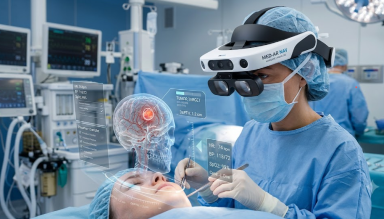

As high-tech AR enters the healthcare sector, a common question arises: “If world-class surgeons can already perform ultra-delicate procedures, why do they need an AR navigation headset equipped with microdisplays?” [1]
The answer is straightforward: even the most seasoned surgeons cannot see through opaque human tissue with the naked eye [2]. Medical instruments have always been essential extensions of surgical practice. Furthermore, during marathon procedures, a surgeon’s cognitive capacity faces physiological limits, while the margin for error under the scalpel remains strictly zero. Technology advances precisely to drive surgical risks as close to zero as possible [3].
The Three Major Pain Points in Modern Surgery
Even renowned international experts must continuously grapple with three fundamental surgical pain points when operating on complex lesions—such as deep skull base tumors, severe spinal deformities, or deep-seated organ lesions [2], [4]:
1. Cognitive Burden of Mental 3D Reconstruction
In traditional operating rooms, a surgeon’s gaze constantly toggles between the surgical field and adjacent 2D MRI or CT monitors. Surgeons must rely entirely on memory and experience to mentally reconstruct two-dimensional cross-sectional slices into a three-dimensional anatomical model, and then map that model onto the physical patient. This constant spatial translation induces severe cognitive fatigue [1], [4].
2. Anatomical Blind Spots in Opaque Tissue
Human organs are opaque. Tiny nerves and blood vessels hidden behind tumors, deep muscle layers, or bone structures remain entirely out of sight before dissection. To expose these concealed structures, surgeons often have to enlarge incisions or over-retract healthy tissue, inevitably increasing postoperative pain and recovery time [2], [5].
3. Physiological Limits of Minimally Invasive Precision
When inserting screws into cervical pedicles, the safe corridor is often a mere 1- 2mm wide. Over hours of intense focus, visual fatigue and imperceptible hand tremors can escalate the risk of surgical complications [6]. Consequently, the number of complex procedures a surgeon can safely perform is physically capped, contributing to a global shortage of specialized surgical care [2].
AR surgical navigation systems are not designed to replace a surgeon’s clinical expertise; rather, they eliminate human physiological blind spots [1]. Powered by high-resolution, high-contrast microdisplays (Micro-OLED), AR technology overlays hidden vascular, neural, and tumor structures directly onto the surgeon’s primary field of view at a 1:1 scale—achieving seamless alignment between brain, hand, and eye [1], [3].
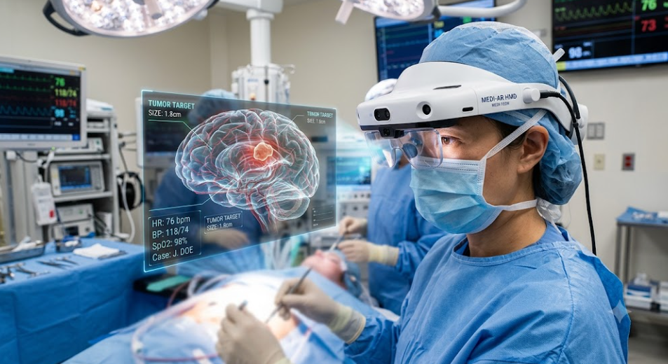
How AR Surgical Navigation Systems Assist in High-Stakes Surgeries: Case Applications
In AR surgical navigation systems, the core value lies not merely in “displaying an image,” but in achieving true x-ray vision and perfect hand-eye alignment [1].
Below are four of the most demanding surgical scenarios where microdisplay overlays and AR navigation provide critical clinical utility:
1. Neurosurgery: Complex Giant Intracranial Aneurysm Clipping and Microvascular Bypass
Intracranial aneurysms represent ticking time bombs in cerebral vasculature, particularly complex aneurysms located at the internal carotid artery or basilar tip [5]. Their walls are paper-thin and under high tension; a minor error can cause catastrophic, jet-like hemorrhaging. In traditional clipping, the aneurysm bulk obscures hair-thin perforating vessels on its dorsal side. These tiny vessels supply blood directly to the brainstem and internal capsule; inadvertently clipping them leads to immediate hemiplegia, persistent vegetative state, or death [5].
- AR Navigation Application: Wearing a high-resolution AR headset integrated with preoperative 3D-CTA/DSA and hemodynamic data, the surgeon sees a translucent 3D aneurysm model and its underlying perforating vessel network precisely superimposed over the physical anatomy at a 1:1 ratio [1]. Pseudo-color dynamic arrows indicate real-time blood flow velocity and direction, eliminating visual blind spots [3]. Furthermore, before deploying a physical titanium clip deep into the brain, the AR interface projects a virtual clip at the aneurysm neck to simulate the optimal trajectory and closure path [5].During temporary arterial occlusion or microvascular bypass, a floating AR HUD displays a real-time brain tissue ischemia countdown timer. Combined with intraoperative fluorescence angiography, it highlights perfusion recovery in distal vessel beds, ensuring no stitch compromises cerebral blood supply [3], [5].
2. Spine Surgery: Upper Cervical Spine and Severe Deformity Pedicle Screw Placement
The osseous pedicle channels in the cervical spine (especially C1/C2) often measure just 3- 5mm in diameter, bounded internally by the spinal cord and externally by the vertebral artery [6]. A medial deviation of just 1\ mm can puncture the spinal cord, causing high quadriplegia; a lateral deviation of 1mm can rupture the vertebral artery, triggering fatal hemorrhage or cerebral infarction [6]. Traditional reliance on landmark palpation and repeated C-arm X-rays involves high radiation exposure and limited precision.
- AR Navigation Application: Looking at the patient’s exposed spine, the surgeon sees a bright green virtual trajectory projected directly onto the vertebra—acting as a precision aiming reticle [6]. Floating telemetry on the HUD displays pitch/yaw angles, real-time drill depth, and remaining bone wall thickness. If the hand drill strays more than 0.5 from the planned trajectory, the AR light axis instantly flashes red to signal a warning, making screw placement vastly safer [1], [6].
3. Hepatobiliary Surgery: Deep Caudate Lobe Tumor Resection & Intrahepatic Vascular Reconstruction
The liver is a highly vascular organ traversed by complex, blood-filled portal, hepatic, and arterial networks [7]. The caudate lobe, nestled in the deepest region of the liver, is tightly sandwiched between the inferior vena cava and the main portal vein. Removing a caudate tumor while preserving adequate hepatic inflow and outflow presents a major challenge, as hidden vessels can easily be severed, leading to hemorrhagic shock [7].
- AR Navigation Application: A 1:1 preoperative 3D CT reconstruction of the intrahepatic vascular tree is projected onto the liver surface. As the surgeon dissects the hepatic parenchyma layer-by-layer with an electrosurgical unit, the AR interface dynamically slices open the semi-transparent 3D liver model, revealing in real time which branch vessel lies just 2 mm beneath the instrument tip [1], [7]. Virtual color staining clearly delineates resection planes for targeted vascular occlusion, preventing collateral damage to healthy liver tissue [7].
4. Orthopedics & Craniomaxillofacial Surgery: Complex Craniomaxillofacial Fracture Reduction & 3D Reconstruction
Severe facial fractures caused by high-impact trauma result in fragmented bone structures and a total loss of anatomical landmarks [8]. Intertwined with complex facial and optic nerves, these fractures make traditional reconstruction reliant on spatial guesswork, frequently leading to postoperative facial asymmetry, enophthalmos, or facial paralysis [8].
- AR Navigation Application: An AR navigation system mirrors 3D data from the patient’s undamaged side and overlays a virtual, semi-transparent skeleton onto the fracture site [8]. As the surgeon manipulates bone fragments with reduction forceps, AR guidance arrows indicate the exact trajectory for anatomical reduction. When the fragment reaches its anatomically correct position, the overlay turns bright blue (“perfect alignment”), dramatically reducing operative time [1], [8].
Technical Requirements for Micro-OLED Displays in High-Stakes Surgery
- Zero-Latency Spatial Alignment: Motion-to-photon latency must be near-zero; the floating AR vascular map must remain locked to physical organs without lag or drift during head movements [1].
- Ultra-High Contrast & High Pixel Density (PPI): In operating rooms with intense surgical spotlights and dim ambient lighting, micro-overlays must render razor-sharp edges and pure luminescence without background glow [1], [3].
- Rigorous Binocular Consistency (Binning & Calibration): Brightness and color temperature across left and right microdisplays must be perfectly matched to eliminate binocular rivalry and prevent visual fatigue during 3–5 hour procedures [1].
Commercial Landscape: Market-Leading AR Surgical Navigation Platforms
The transition of AR surgical navigation from academic research to clinical standard-of-care is already underway, spearheaded by pioneering medical technology companies:
-
Augmedics (xvision Spine System®): As the first FDA-cleared AR guidance system for spine surgery, xvision utilizes a proprietary head-mounted display (HMD) with built-in optical tracking [9]. It projects 3D trajectory lines and anatomical data directly onto the surgeon’s retina, allowing spine surgeons to visualize internal spinal structures through the patient’s skin without looking away at external monitors [6], [9].
-
Brainlab (SyncAR & Mixed Reality Navigation): Partnering with leading optical and head-mounted display developers, Brainlab integrates patient-specific 3D MRI/CT reconstructions directly into the surgical field and microscope optics [10]. In neurosurgery, SyncAR allows clinicians to overlay intracranial vascular trees and tumor margins in real-time, delivering millimeter-level tracking accuracy during complex cranial procedures [4], [10].
-
Surgical Theater (SuRgicalAR™): Built on flight-simulation algorithm technology, SuRgicalAR™ converts standard 2D patient scans into dynamic, 3D holographic models [11]. Cleared for intraoperative AR visualization, it enables neurosurgeons to conduct virtual “flight-throughs” during preoperative planning and overlay real-time vascular dynamics onto the surgical field during aneurysm clipping [5], [11].
-
Novarad (OpenSight™ AR Surgical System): An FDA-cleared AR visualization system designed for preoperative mapping and intraoperative guidance [12]. OpenSight™ projects interactive 3D holograms onto the patient’s body surface, assisting orthopedic and traumatology surgeons in precise instrument alignment and complex fracture reduction [8], [12].
References:
[1] Omdia Research. Near Eye Display for XR Application Report & Display Dynamics Tracker, 2024–2026.
[2] IEEE Xplore. IEEE Transactions on Biomedical Engineering – Augmented Reality Navigation in Minimally Invasive Surgery, 2024–2025.
[3] Display Daily. XR in Healthcare and Specialty Markets: Near-Eye Displays in Surgical and Assistive Applications, 2025.
[4] Journal of Neurosurgery. Augmented Reality and Head-Mounted Displays in Complex Cranial Base Surgery, 2024–2025.
[5] World Neurosurgery. Intraoperative 3D Holographic Navigation in Intracranial Aneurysm Clipping, 2025.
[6] Spine Journal. Accuracy and Safety of Pedicle Screw Placement Using Augmented Reality Surgical Navigation, 2024–2026.
[7] Annals of Surgical Oncology. Augmented Reality-Guided Hepatectomy for Deep-Seated Liver Tumors, 2025.
[8] Journal of Cranio-Maxillofacial Surgery. 3D Mirroring and AR Overlay in Complex Maxillofacial Reconstruction, 2024–2025.
[9] Spine Spine Journal / FDA Medical Device Database. Augmedics xvision Spine System (XVS) 510(k) Clearance & Clinical Efficacy Study, 2023–2025.
[10] Neurosurgical Focus. Evaluation of Brainlab Mixed Reality and SyncAR Platform in Complex Skull Base and Vascular Neurosurgery, 2024–2025.
[11] Operative Neurosurgery. Intraoperative Augmented Reality via SuRgicalAR Platform for Intracranial Aneurysm and Vascular Lesions, 2024.
[12] Journal of Digital Imaging. FDA-Cleared Augmented Reality Guidance: Clinical Utility of the Novarad OpenSight Platform in Surgical Traumatology, 2024.
Disclaimer
- Informational Purpose Only: This document is intended solely for academic discussion, biomedical engineering evaluation, and technical technology analysis. It does not constitute medical advice, clinical guidelines, or diagnostic recommendations.
- Regulatory Compliance: AR surgical navigation systems and microdisplay hardware discussed herein must comply with applicable regulatory frameworks (e.g., FDA 510(k), CE-MDR) and be operated under approved clinical protocols by qualified medical professionals.
- Intellectual Property: All referenced journals, third-party reports, and institutional trademarks belong to their respective copyright holders. Their inclusion serves strictly for objective scientific discussion.